By Harold (Fritz) Moritz
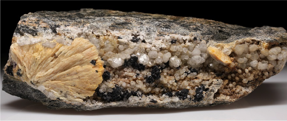
Specimen 1367 - 21 x 6.5 cm. H. Moritz photo.
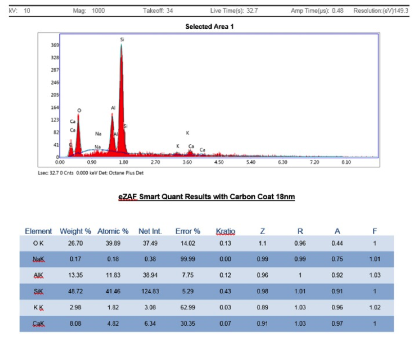
Recently I submitted samples of heulandite subgroup crystals from several places around Connecticut for analyses. Micromounters New England will perform a limited number of scanning electron microscope – energy dispersive spectroscopy (SEM-EDS) analyses for members such as me. SEM-EDS provides elemental chemistry on extremely small samples except for the very light elements H, He, B, Be and Li.
The goals of this project were to better determine the particular species of heulandite on the specific samples and from that information hopefully extend the species identifications to unanalyzed specimens from the same localities, and perhaps throughout the state. Consequently, 3 samples were submitted from the very prolific O&G basalt quarry locality and 3 samples from veins and faults hosted by metamorphic rocks. All were initially identified based on their crystal habit and physical properties
Originally described as one mineral in 1822, the heulandite subgroup of zeolite minerals has been divided into 5 similar-looking species - chabazite-Ba, chabazite-Ca, chabazite-K, chabazite-Na, chabazite-Sr, having the general formula (A)5(Si27Al9)O72 · 26H2O, where the A site can be occupied by Ba, Ca, K, Na, and/or Sr. The particular species depends on the relative proportion of the elements in the A site. In general, according to mindat.org, chabazite-Ca appears to be by far the most common species. However, some of the disparity may be due to a lack of analyses. But with few if any published modern analyses of local samples, anything could be found locally.
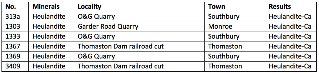
Below is a table of the samples from my collection followed by a discussion of the results.
Heulandite crystals occur in Connecticut only at a few places in the basalt flows (trap rock) where there is significant mineralization in gas vesicles and fractures. These are primarily the two O&G quarries in Woodbury (No. 1) and on the Woodbury/Southbury line (No. 2). In this geo-environment it typically occurs with datolite, pumpellyite, calcite, quartz, babingtonite, apophyllite-K, and other zeolite group minerals (stilbite, chabazite, analcime, natrolite). Many attractive specimens have been saved from these localities.
Heulandite can also rarely occur in fractures in metamorphic rocks with other zeolite minerals, typically stilbite. It seems to be the second most common zeolite in these fractures after stilbite, but they are usually very small and easily missed, so carefully look specimens over if stilbite is obvious. The Garder Road Quarry crystal was found hiding in with a myriad calcite crystals. But the Thomaston Dam railroad cut yielded many obvious heulandite crystals, so 2 samples were included from here.
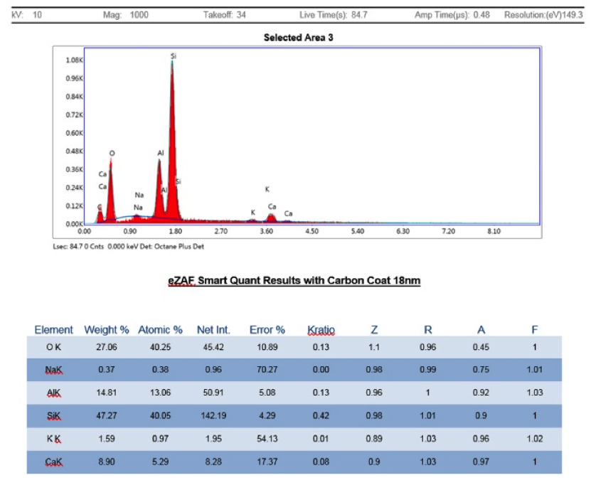
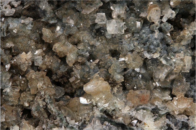
All of the SEM-EDS results shown below found the heulandite-Ca species, with lesser K and Na also detected, but not enough to change the species. No obvious difference in the chemistry between crystals found in basalt and those found in schist or gneiss. I think it is safe to conclude that heulandite found elsewhere in the state should belong to the chabazite-Ca species, unless it is identified at a new locality that has a very different mineral-forming geo-environment than the ones above. In that case, a full analysis should be done.
Specimen 313
Peach colored chabazite-Ca rhombs on white quartz alteration of chalcedony that surrounded dissolved tabular anhydrite molds. A few pearly, colorless heulandite-Ca crystals are also present at lower center between two vertical rows of chabazite-Ca.
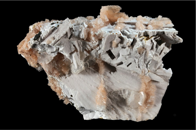
Specimen is 7 x 5 cm. H. Moritz photo.
Specimen 1303
Brown heulandite-Ca crystal surrounded by calcite crystals on peachy albite, in a fracture in gneiss. The little bit of Mg in the EDS spectrum likely results from whatever inclusions are coloring this crystal.
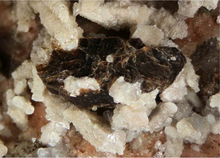
FOV 12.8 mm. H. Moritz photo.
Specimen 1333
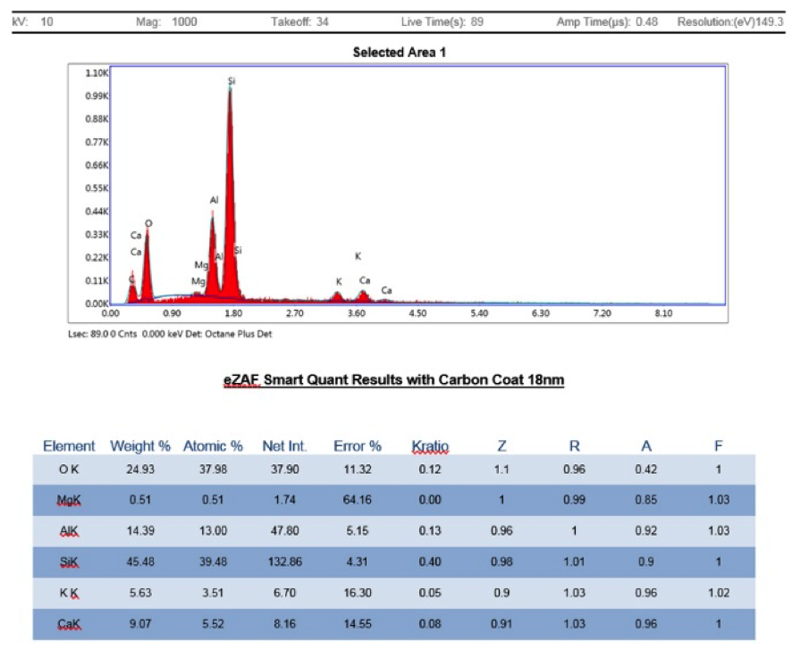
Tan heulandite-Ca crystals with white calcite in a fracture in basalt.
Field of view 2.8 cm. H. Moritz photo.
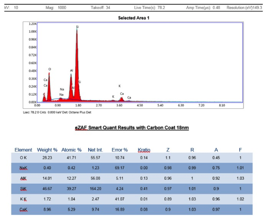
Specimen 1367
Typical mineralized fracture in schist from Thomaston Dam railroad cut, showing tan, partial stilbite “fan” of crystals at left, milky quartz with black sphalerite, and a tan, partial heulandite-Ca crystal at right from which the sample was taken.
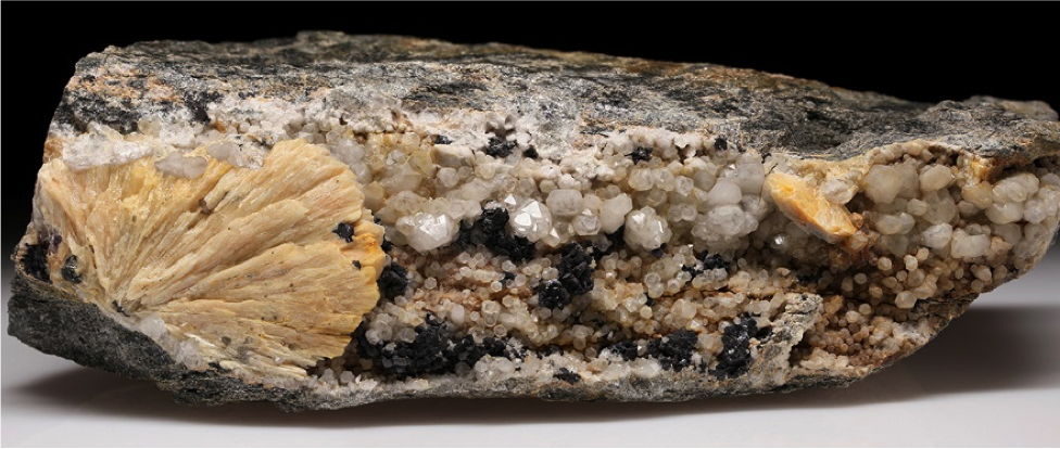
FOV 21 x 6.5 cm. H. Moritz photo.
Specimen 1369
Tan heulandite-Ca crystals on a bed of calcite, in a fracture in basalt.
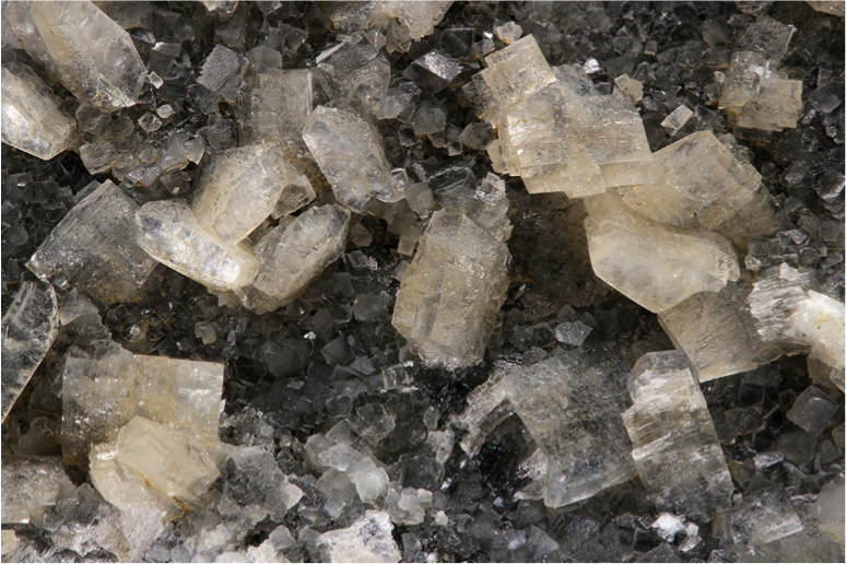
Field of view 2.2 cm. H. Moritz photo.
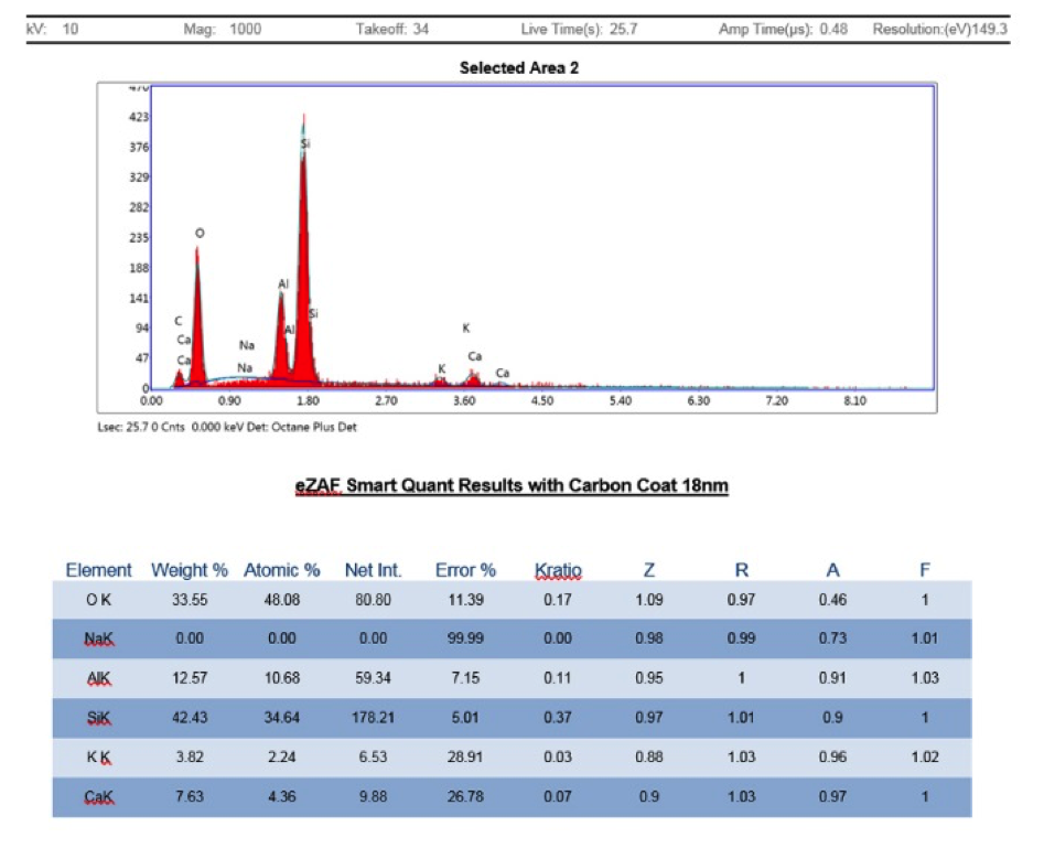
Specimen 3409
A fracture in schist with yellow stilbite, white calcite, and pale tan, finer-grained heulandite sprinkled with pyrite crystals.
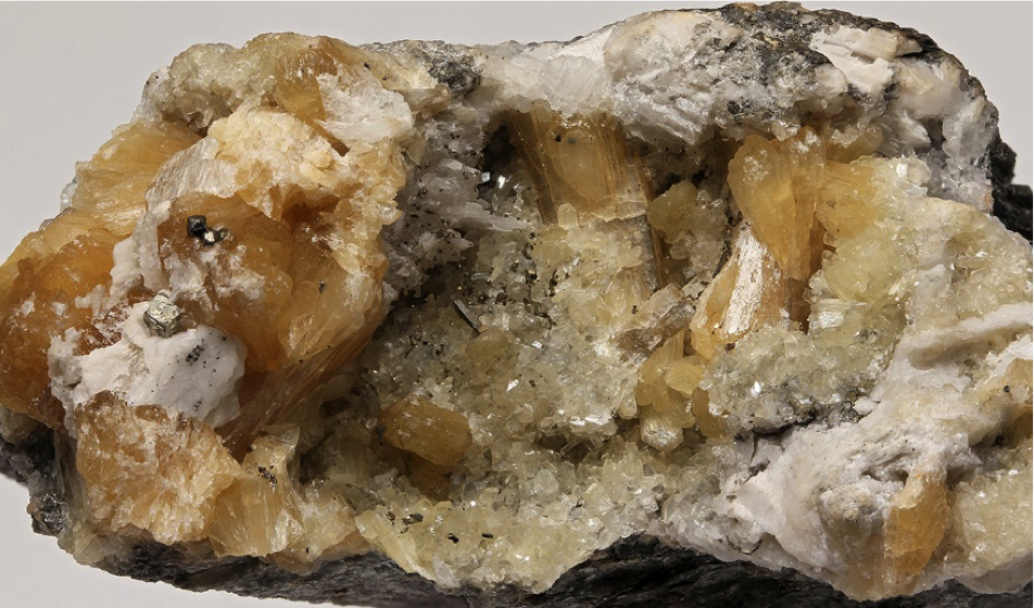
Specimen is 6 x 3.5 cm. H. Moritz photo.