|
by Harold (Fritz) Moritz
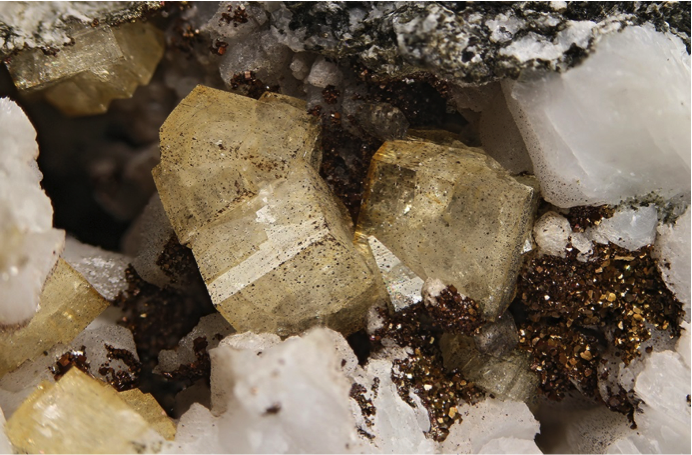
Specimen MP-03. Field of view 22mm. Mike Polletta specimen. H. Moritz photo.
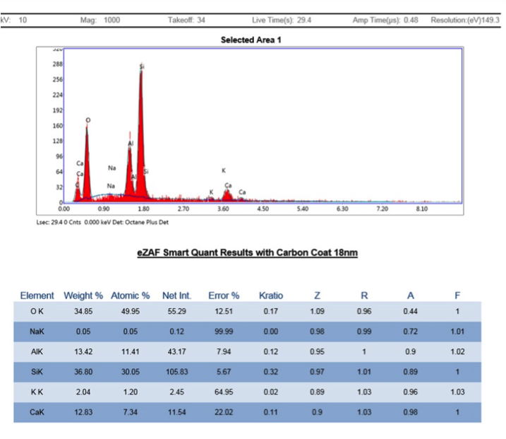
Recently I submitted samples of chabazite group crystals from several places around Connecticut for analyses. Micromounters New England will perform a limited number of scanning electron microscope – energy dispersive spectroscopy (SEM-EDS) analyses for members such as me. SEM-EDS provides elemental chemistry on extremely small samples except for the very light elements H, He, B, Be and Li.
The goals of this project were to better determine the particular species of chabazite on the specific samples and from that information hopefully extend the species identifications to unanalyzed specimens from the same localities, and perhaps throughout the state. Consequently, at least one sample was selected from most known localities with 2 samples submitted from the very prolific O&G locality.
Originally described as one mineral in 1788, the chabazite group of zeolite minerals has been divided into 5 similar-looking species - chabazite-Ca, chabazite-K, chabazite-Mg, chabazite-Na, chabazite-Sr, with most of them having the general formula (A)2[Al2Si4O12]2 · 12H2O, where the A site can be occupied by Ca, K2, Na2, and/or Sr. The particular species depends on the relative proportion of the element in the A site. Chabazite-Mg has a slightly different formula. In general, according to mindat.org, chabazite-Ca appears to be by far the most common species, with chabazite-Na, chabazite-K relatively uncommon, and chabazite Mg and chabazite-Sr very rare. However, some of the disparity may be due to a lack of analyses. But with few if any published modern analyses of local samples, anything could be found.
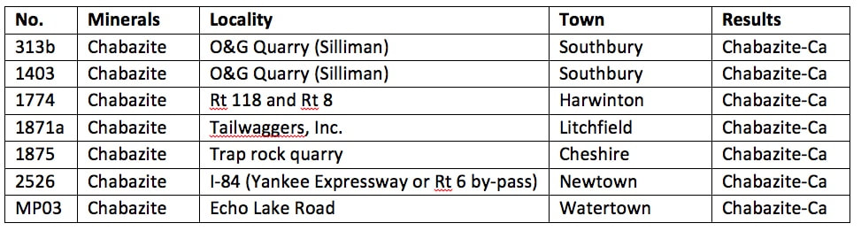
Below is a table of the samples (most from my collection except MP03) followed by a discussion of the results.
Chabazite crystals occur in Connecticut only at a few places in the basalt flows (trap rock) where there is significant mineralization in gas vesicles and fractures. These are primarily the two O&G quarries in Woodbury (No. 1) and on the Woodbury/Southbury line (No. 2). It is also less commonly found at the quarry at Reed’s Gap on the Durham/Wallingford line and at the old Cheshire Quarry. In this geo-environment it typically occurs with pumpellyite, calcite, quartz, babingtonite, apophyllite-K, and other zeolite group minerals (stilbite, heulandite, analcime, natrolite). Attractive specimens have been saved from these localities.
Chabazite can also rarely occur in fractures in metamorphic rocks with other zeolite minerals, typically stilbite. Several places have yielded many crystals, particularly the old Route 6 bypass in Newtown that became part of I-84, at road cuts in Harwinton and Watertown, and at outcrops at the Tailwaggers, Inc. property in Litchfield.
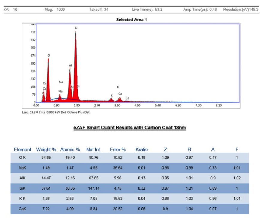
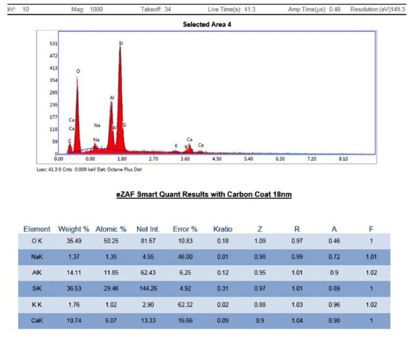
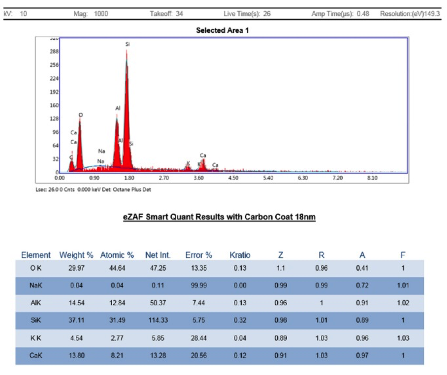
All of the SEM-EDS results shown below found the chabazite-Ca species, with K and Na also detected in most, but not enough to change the species. Interestingly, the specimens from trap rock geo-environments (313, 1403, 1873) included, according to the SEM-EDS operator Peter Cristofono, “minor K, and trace Na” while those from fractures in metamorphic rocks included “significant K, and minor Na” or Mg (1774, 1871, 2526, MP03). The latter tend to have a yellow-orange color, probably from a tiny amount of Fe, too low to show up in the EDS spectra. Thus there is some apparently consistent minor difference in the mineral’s chemistry between these two geo-environments. In any case, I think it is reasonable to conclude that any chabazite found elsewhere in the state should belong to the chabazite-Ca species, unless it is identified at a new locality that has a very different mineral-forming geo-environment than the ones above. In that case, a full analysis should be done.
Specimen 313
Rhombohedral chabazite-Ca crystals on quartz filling in gaps between dissolved tabular anhydrite molds.
Field of view 17mm. H. Moritz photo.
Specimen 1403
Clusters of dark, pseudocubic chabazite sit next to colorless, pearly heulandite (center and right) and frosty calcite (lower left) in a spray of tabular voids left by dissolved anhydrite in white chalcedony agate.
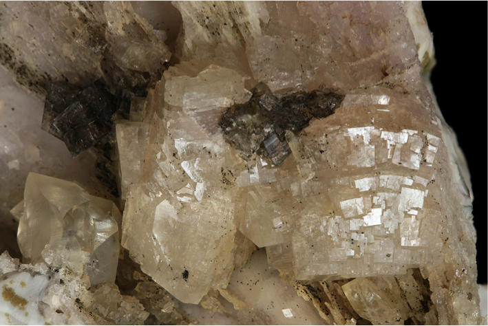
Field of view 26mm. H. Moritz photo.
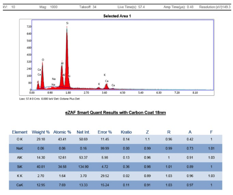
Specimen 1774
Chabazite-Ca crystals on a fracture surface in schist, very surprisingly these crystals fluorescence green under short-wave ultraviolet light.
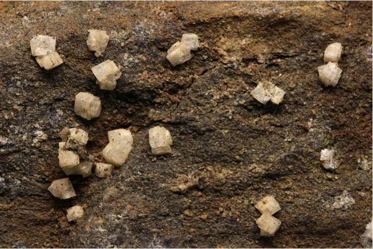
Field of view 4.5 cm. H. Moritz photo.
Specimen 1871
Yellow-orange chabazite-Ca and white stilbite (upper right edge) crystals in a fracture in amphibolite.
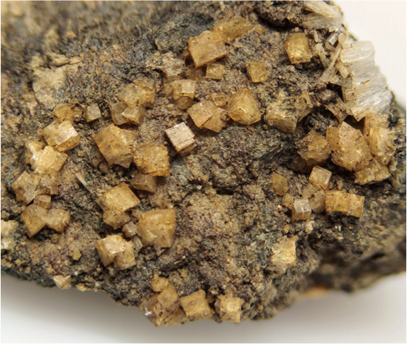
Field of view 20mm. H. Moritz photo.
Specimen 1875
Complex, multiple interpenetration twinned chabazite crystal on calcite, typical of the one from the same piece used for the analysis. All of them are microscopic.
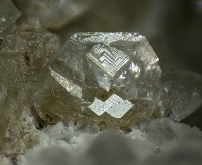
Field of view 2.69mm. H. Moritz photo.
Specimen 2526
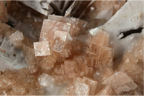
Yellow-orange rhombohedral chabazite-Ca crystals with white calcite on annite gneiss.
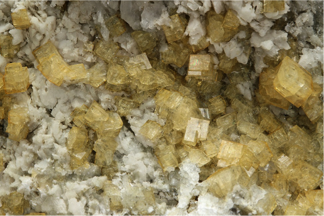
Field of view 2.5cm. H. Moritz photo. 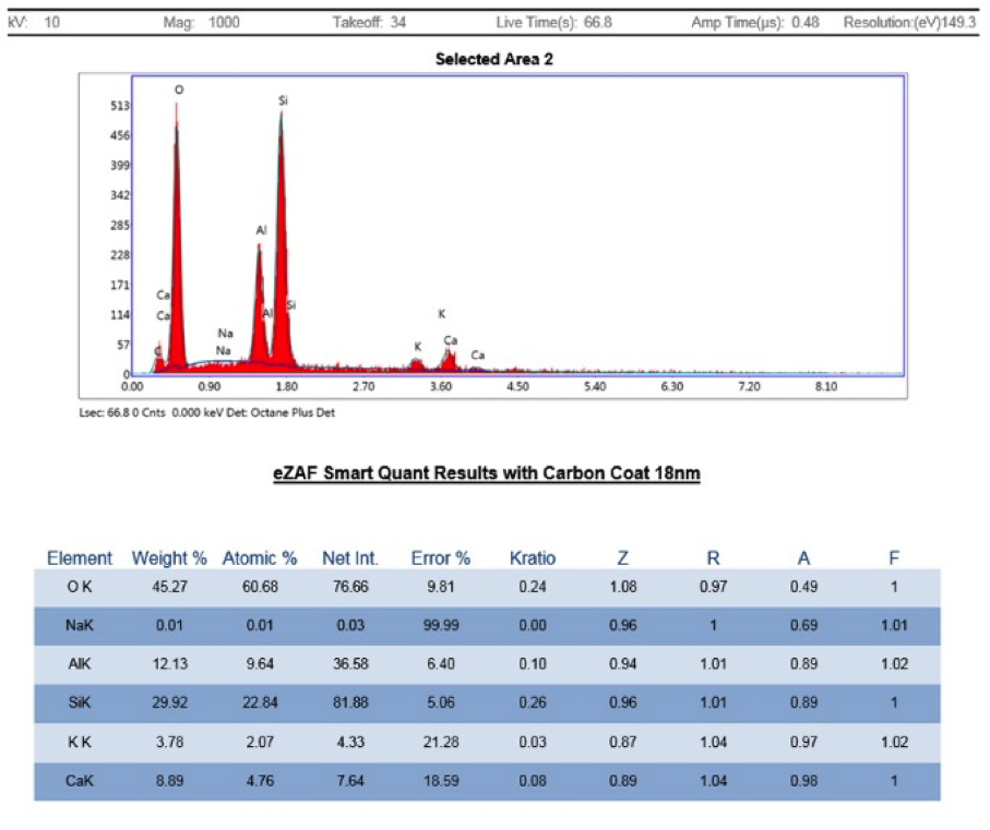
Specimen MP-03
Yellow-orange chabazite-Ca with pyrite and white calcite in a fracture in granofels.
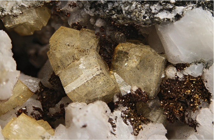
Field of view 22mm. Mike Polletta specimen. H. Moritz photo.
|

